Electron microscopes have long been indispensable tools in scientific research, offering unparalleled resolution and magnification capabilities. However, current electron microscopy technologies face significant limitations, including high cost, large size, strong radiation damage to samples through interaction with the electron beam, and the need for cryogenic temperatures. A research team from City University of Hong Kong (CityUHK) is working on a new quantum electron microscope (QEM) to eliminate interaction between the electron beam and sample. At this stage, the team is using partial key components of QEM to design a compact hybrid transmission and scanning electron microscope that can operate at room temperature, ushering in a new era for electron microscopes. The CityUHK team plans to manufacture and commercialise this groundbreaking innovation within three years.
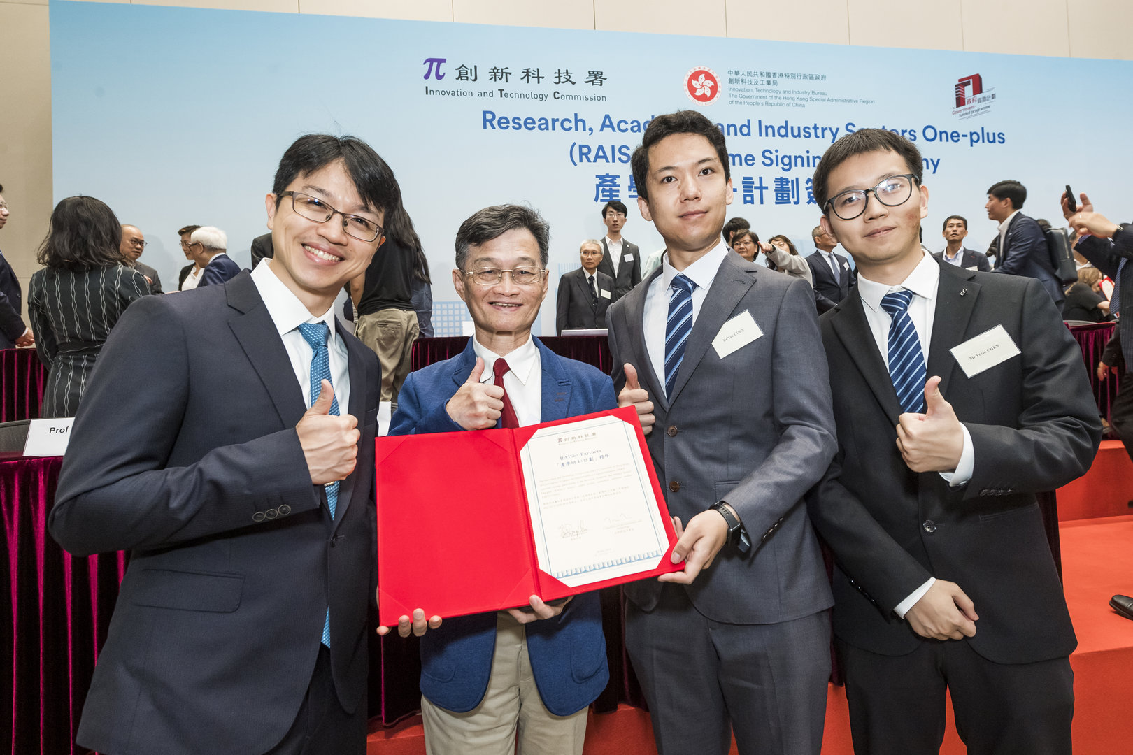
The project, titled “Commercialization of Pulse Hollow Cone Hybrid TEM/SEM”, was led by Professor Chen Fu-rong, Chair Professor in the Department of Materials Science and Engineering at CityUHK. It recently secured funding from the Research, Academic and Industry Sectors One-plus Scheme (RAISe+ Scheme), launched by the Innovation and Technology Commission to unleash the potential of local universities in the transformation and commercialisation of their research.

Transmission electron microscopes (TEMs) and scanning electron microscopes (SEMs) are essential tools in many modern scientific research projects. They provide the high-resolution imaging necessary to study the intricate details of various materials, ranging from biological specimens to nanostructures.
However, the high-energy electron beams that the TEMs and SEMs use can cause significant radiation damage to delicate biological samples. In the structural biology field, cryo-transmission electron microscopy (cryo-TEM) is used to minimise radiation damage by placing proteins in a layer of vitreous ice. However, the ice layer introduces imaging noise, hampering resolution.
In response to these challenges, Professor Chen and his research team at CityUHK designed the Pulse Electron Hollow Cone Illumination Hybrid TEM/SEM, based on technology developed in CityU Shenzhen Futian Research Institute (currently called CityU Matter Research Institute (Futian)).
This innovative system addresses the limitations of existing electron microscopes in several ways. First, its pulse electron source reduces the radiation damage of soft material samples, which is particularly crucial for biological samples. Second, the hollow cone illumination offers about four times greater image contrast than that of bright field images in transmission electron mode, enabling more detailed imaging of the samples. The team will also use their previously developed chromatic and spherical aberration (CS/SS) correctors to further improve spatial resolution.
The new hybrid TEM/SEM system is more cost-effective than conventional TEMs and SEMs. Operating at a low voltage range of 15–30 keV, it can perform 3D protein molecule reconstruction and nano-material investigation at room temperature, surpassing the capabilities of cryo-EM.
The team demonstrated that the system’s high-resolution imaging capabilities in various applications, including imaging metal contacts on printed circuit boards, nanoparticles and other biological samples, achieved a super-high surface resolution better than 10 nm.
Ultimately, the new electron microscope is expected to be operated in transmission mode to observe the 3D structure of proteins and molecules, as well as in scanning mode to observe nano-materials and for semiconductor and chip detection.
“Compared to existing desktop SEM systems, our pulse electron hollow cone system offers excellent SEM imaging quality, comparable to that of the best desktop systems,” said Professor Chen. “There are no equivalent electron microscopes in the market that provide the quality of our system. The unique capability of our pulse hollow cone illumination, allowing 3D protein reconstruction via TEM mode, is not available in any existing desktop SEM.”
“With the Raise+ funding, as well as support from our industry partner, we are planning to establish a mass production line for the commercialisation of these innovative, powerful and compact hybrid electron microscopes within three years,” he added.
Professor Chen has long been engaged in the research field of materials science and electron microscopes. In April 2023, he and his team created a time-resolved electron microscope integrated with both scanning and transmission electron microscope modes in a compact format, becoming the first university-based research team to achieve such a breakthrough.